
A hip ultrasound is a painless and safe test that makes images of a baby’s hips using sound waves, done to babies of up to around six months.
The doctor uses an ultrasound machine that sends sound waves to the hip area and records images on a computer during the procedure.
The images are usually black and white, and they help show the hip structures, including the femoral head (thigh bone) and acetabulum (its socket) located in the pelvic bone. It helps see if there are any structural issues with a baby’s hips.
Why It’s Done
Doctors will perform a hip ultrasound in babies if they suspect that the baby has developmental dysplasia of the hip (DDH). This is a deformity that occurs in babies before, during, or a few weeks or months after birth.
In a normal healthy hip, the femoral head comfortably rests in the acetabulum, but in children with DDH, the bone moves back and forth. The bone might pop out of the socket in some serious cases, but doctors can put it back by applying pressure. It may be impossible to put back the bone into the socket in severe cases.

Source: onlinelibrary.wiley.com
Some of the things that may cause DDH include:
- Abnormal baby positions in the womb
- Babies being cramped up inside the uterus mostly because of a decrease in amniotic fluid
- Family history of the disorder
Procedure
When taking your baby for an ultrasound, you do not have to do major preparations. You only have to tell the technician if they are under any medications.
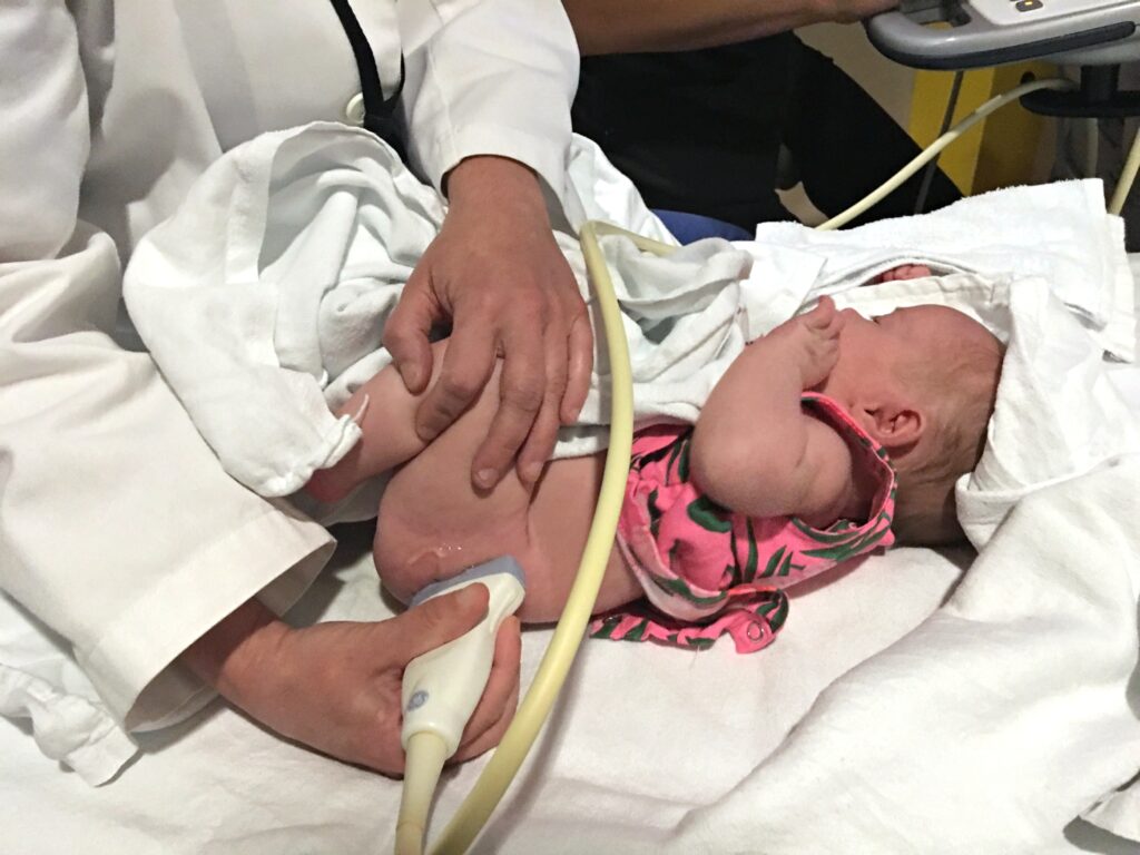
The technician (sonographer) usually performs the ultrasound in a radiology center or the hospital’s radiology department. They also ensure the room is dark so that they can see the images clearly on the computer.
The sonographer will ask you to undress the baby and remove the diaper partially, and then they will place your baby on the ultrasound table on their side or back.
They then spread a clear gel on the targeted area of the hip. The gel helps in the proper transition of sound waves. The sonographer then moves a transducer over the gel. That is a small handheld wand that emits high-frequency sound waves.
The computer then measures how the waves bounce back from your baby’s body and change them into images. Sometimes a doctor will come in before the end of the session to examine the baby and take more photos.
The procedure is usually painless with only slight pressure on the skin. The sonographer finishes by wiping off the gel, and you can dress your baby.
The procedure normally takes 20 minutes but may take longer depending on the complexity of the issue or the state of your child. Ultrasound exams are sensitive to motions, and if your child keeps moving, it could give wrong readings and last longer.
Therefore, doctors advise that you try your best to calm your baby throughout the procedure and, if possible, bring toys, games, books, or music to keep them distracted. Sometimes the exam room may have a television where you can put on your child’s favorite channel.
What To Expect After The Procedure
Source: budgetsavvydiva.com
A radiologist (a doctor who specializes in reading and interpreting ultrasound and x-ray images) will read and interpret the images of your child’s ultrasound. Results for hip ultrasound in babies take around one or two days to come in.
There are no risks or side effects related to ultrasound in babies because there is no radiation in the procedure.